BTSC Lab Assistant Biology Lab: Microscopy and Specimen Handling Guide
Build an exam-ready microscopy workflow through a labelled onion wet mount, a checked cell-size estimate, safe specimen handling, and six common errors.
KnowledgeGate Team
Exam prep & CS education
Updated 14 Aug 20265 min read
Microscopy questions mix instrument parts, focusing order, slide preparation, observation and safe handling into one sequence, and one missed step in that chain costs the whole answer. A single onion-epidermis wet mount and one field-of-view calculation carry every part of it. Marks, question counts and the topics actually set for your stream come from the applicable notice on the official BTSC website. The rest of this exam’s subject posts sit in the BTSC Lab Assistant blog hub.
1. Define the microscopy and specimen-handling task
Treat the topic as a chain: identify the part, prepare and label the specimen, focus from low to high power, record, estimate size, then clean and dispose according to the lab SOP.
Laboratory practice itself does not change from one notice to the next, so it is worth learning once and keeping in your notes. Syllabus inclusion, question count, marks and negative marking do change, and those come only from the applicable BTSC notice. Revise the chain with this five-column grid:
Step | Instrument or material | Action | Expected observation | Error to avoid |
|---|---|---|---|---|
Identify | Microscope | Name each part | Correct part-function match | Guessing from shape |
Prepare | Slide and specimen | Label, mount and cover | Thin, traceable preparation | Fold or missing label |
Focus | Objectives and knobs | Work from low to high power | Centred, clear image | Coarse focus at 40x |
Record | Notebook | Note code and observation | Traceable result | Unlinked notes |
Finish | Lens paper and disposal area | Clean and segregate | Safe bench | Mixed waste |
The measurement-error arithmetic and the general bench-safety rules sitting behind this grid are worked out for this exam in BTSC Lab Assistant Units, Measurement Error and Lab Safety.
2. Know the microscope before touching the slide
Group parts by function. Eyepiece and objectives magnify; stage and clips support the slide; condenser and iris diaphragm control illumination; coarse and fine knobs focus; the revolving nosepiece changes objectives.
With a 10x eyepiece, total magnification is:
With the 4x objective:
10 x 4 = 40x.With the 10x objective:
10 x 10 = 100x.With the 40x objective:
10 x 40 = 400x.
Begin at 4x. Centre the specimen, bring it up with the coarse knob, then open the iris until the field is evenly lit without glare. Move to 10x and then 40x. At 40x, use fine adjustment only. Never put immersion oil on a dry 40x objective. Clean optics only with lens paper.
3. Worked practical: prepare an onion-epidermis wet mount
Label a clean slide ON-1. With forceps, place one thin 5 mm by 5 mm onion epidermal peel flat in a small water drop. Lower an 18 mm by 18 mm coverslip edge-first at about 45 degrees, then blot only excess liquid at its edge.
If the supervised procedure includes staining, put one small iodine drop at one edge and draw it across with absorbent paper at the opposite edge. Do not flood the slide. Keep droppers separate and never return excess reagent to stock.
At low power, find one thin layer. At 100x total magnification, centre the brick-like cells and visible cell walls. Move to 400x only after centring. Do not expect every organelle to be visible with an ordinary school microscope.
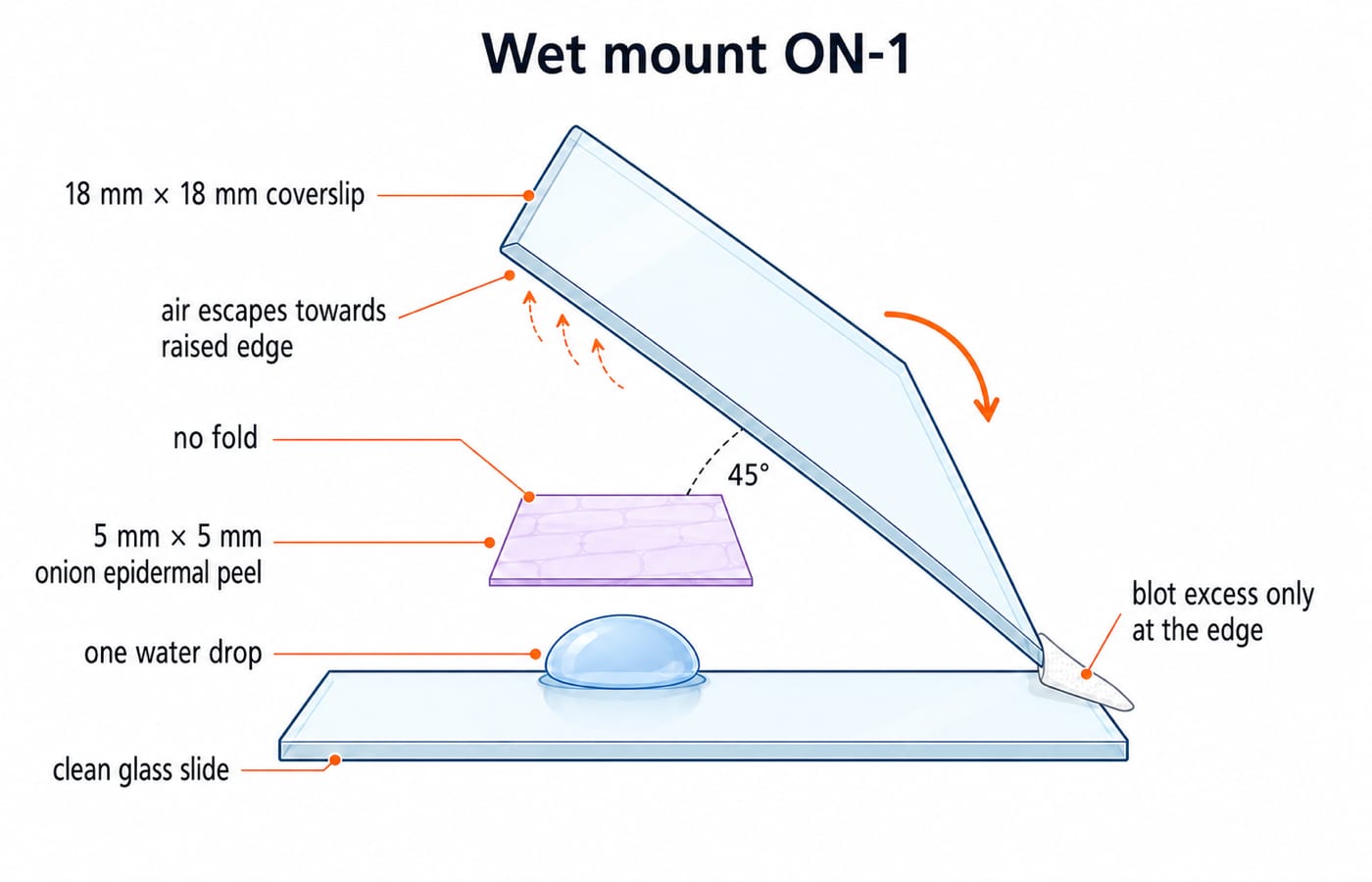
An unlabelled or leaking preparation goes back to the bench. Remake it and label the slide before you look, rather than guessing what is on it.
4. Worked numerical: magnification, field of view and cell size
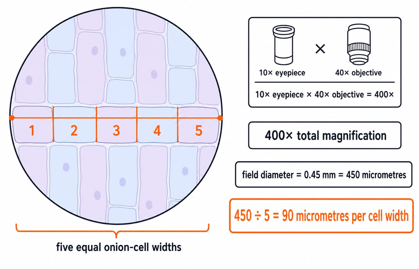
With a 10x eyepiece and 40x objective, total magnification is:
10 x 40 = 400x
Suppose field diameter at the 10x objective is 1.8 mm. On the same microscope it is inversely proportional to objective magnification, so at 40x:
1.8 mm x 10 / 40 = 0.45 mm
Since 1 mm is 1,000 micrometres:
0.45 mm x 1,000 = 450 micrometres
If five equal-looking onion-cell widths fit across the field:
450 micrometres / 5 = 90 micrometres per cell width
This estimate assumes five equal-looking widths. It does not make every onion cell exactly 90 micrometres wide.
5. Handle specimens without losing identity or contaminating the bench
For traceability, label first, open one specimen, use a clean tool, close the container, record the observation, then use the correct return or disposal area. Record the specimen code, source or type, preparation or stain, date, and observer initials where the SOP requires them.
Do not collect a cheek smear or handle blood, sputum or unknown material outside a supervised lab. Use prepared teaching slides unless the lab’s own biosafety procedure authorises collection and disposal. Wear required PPE, put sharps in the designated container, and never mouth-pipette. Follow the institution’s named disinfectant, dilution and contact time. There is no universal concentration.
6. Diagnose the six errors exam questions hide
No image after changing objective: the specimen was not centred. Return to 4x, re-centre and refocus.
Dark field: illumination or the iris is poorly adjusted. Reset the light and condenser.
Blurred image at 40x: coarse focusing or a dirty lens is likely. Use fine adjustment and lens paper.
Many round, clear circles: air is trapped. Remake the mount and lower the coverslip at 45 degrees.
Overlapping cells: the peel is folded or too thick. Use one thin, flat layer.
Observation cannot be traced: the slide was not labelled. Stop, and restore identity only from a valid record.
Ordinary tissue can scratch an optical surface. Pressing a coverslip can break the glass or distort the specimen. Both are traps, not shortcuts.
7. The short version: convert the workflow into recall
Check four answers: 10x eyepiece with 40x objective gives 400x; at 40x use fine adjustment; five widths across 450 micrometres give 90 micrometres per width; lowering the coverslip edge-first at about 45 degrees reduces bubbles.
Use one 20-minute loop: five minutes each to label the microscope, recall the wet-mount sequence, repeat the 1.8 mm to 0.45 mm calculation, and correct the six errors.
Continue from the BTSC Lab Assistant category. KnowledgeGate’s BTSC Lab Assistant complete course and BTSC Lab Assistant test series are built for the Computer Science stream. If you are sitting the Biology paper, use them for the computer section only and keep your microscopy revision on the workflow above.
Keep learning
BTSC Lab Assistant Physics Lab: Instruments, Readings and Error Calculations
Learn a fixed method for selecting lab instruments, reading their scales, correcting zero error and reporting calculated results with sensible uncertainty.
Updated 16 Aug 20266 min readBTSC Lab Assistant
BTSC Lab Assistant Job Profile: Records, Equipment Care and Daily Lab Workflow
Build a realistic CS/IT Laboratory Assistant role model around readiness checks, auditable registers, safe fault handling and evidence-based handover.
Updated 12 Aug 20265 min readBTSC Lab Assistant
BTSC Lab Assistant PYQ Analysis: Sort Common and Trade Topics
Turn a mixed PYQ score into a useful repair plan. This method separates common and trade topics, codes each miss and assigns revision time from your own errors.
Updated 10 Aug 20266 min readBTSC Lab Assistant

BTSC Lab Assistant Chemistry: Solutions, Titration and Lab Safety
Connect solution preparation to acid-base titration through a checked HCl and NaOH example. Learn the apparatus, calculations, error directions and safe lab sequence.
Updated 7 Aug 20266 min readBTSC Lab Assistant